Home » Posts tagged 'Brain'
Tag Archives: Brain
If the NHS is like a human brain, we may have to think about reconfigurations a bit differently
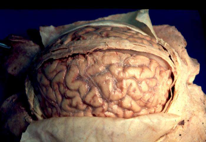
The human brain is probably the most complex organ of the body, and comparing it to the National Health Service may give us conceptual hints as to how we approach reconfigurations.
The brain cannot undergo limitless growth in response to increasing demand, which may or may not increase as a person gets older.
This is shown for example by the fact that it’s contained within a skull. It has an outer covering which encases it. This is known as the ‘blood brain barrier’ regulating traffic between the body and the brain, analogous to the regulators.
Seeing what is happening from output this barrier can be a good marker of the quality of activities within the brain itself.
The brain makes decisions, plans ahead, makes decisions, has a working memory, can learn, has memory for events, and is able to perceive aspects of the world around it.
Also bits of it can suffer from a build-up of toxic metabolites, which may be kept hidden.
It needs a brainstem to survive, which for example contains centres that control our breath. This brainstem also connects the brain to the rest of the body.
We of course need lungs to breathe. In the same way, the the NHS needs the rest of the economy to survive. And we can’t absolutely divorce what is happening in the NHS from the rest of what is happening in the UK, in much the same way the brain cannot be thought to be separate from lungs. There’s an interdependency.
The human brain is incredibly metabolically active. Without being given adequate resources, the whole thing will simply die.
The body needs to work too. For example, if we can’t breath, we don’t get the oxygen to run the brain. If there is insufficient monies to run the NHS, of the order of billions, it will simply fail.
No-one really understands the complexity of the brain, in a similar way to not many people understand how the NHS works as a whole. There are thought to be 100 billion brain cells, in various proportions of the types of brain cells.
With so many brain cells, and connections between them, it’s inevitable that some connections (and even brain cells) will become “redundant” with time.
The number of each category of brain cells doesn’t matter as such as long as the whole brain functions well. However, some people worry that certain types of brain cells, or entities in the NHS, consume too much energy and are not particularly efficient. This is of course of concern if you’re thinking about having enough energy for the brain as a whole.
It’s likely that no one part of the brain fulfils a certain function in the brain, such as decision making. It’s likely that different parts of the brain work in synchrony to fulfil these functions. However, some parts of the brain may be more important than others for producing certain brain functions.
This is possibly helpful to think about what happens when some parts of the brain fail.
Some parts of the NHS may be running a deficit in a moribund way before they enter an outright failure regime.
In the same way, it could be that some parts of the brain don’t have enough oxygen – maybe insufficient money or spending too much money, and so forth. Lack of oxygen can produce a stroke.
The question is what happens when a part of the brain has a stroke. Somebody has to ascertain that that part of the brain has no residual function left.
Who makes that diagnosis, that there’s no function left, and no amount of oxygen will make that brain part work, is bound to be controversial, as to the subsequent management step of allowing that brain part to go peacefully out of action.
A problem obviously arises if it’s decided that that ‘out of action’ brain part goes into managed decline too quickly.
It could be that that part of the brain is actually critical for a function from the brain. This could be the case for a particular entity in the NHS itself too.
If a part of the brain is truly dead, the rest of a network of other parts of the brain could be recruited to fulfil that function. Such plasticity is known to a limited degree in the adult nervous system, but is much easier in the developing nervous system.
Brains which are old depend on activity in parts of the brain to determine where the connections are strongest and best made.
This is fundamentally the ‘issue’ for socialists who think that activity can determine some NHS services going into ‘managed decline’ through a mercenary neo-Darwinian ‘survival of the fitness’ process.
A problem with Lewisham, originally, was that it appeared that bits of the brain, to overegg this analogy, were being shut down because other parts of the brain had failed. This is of course counterintuitive.
It could be through proper planning can we can work out how to reconfigure networks so that the brain is still able to provide all its functions.
A major problem with recent legislation is bits of the brain can now be shut down without proper analysis,
Another major problem is that there is enormous public distrust, so that people are uncertain that the brain will be given enough oxygen to carry out its functions.
But on a happier note, the brain is a bit unpredictable. It is a remarkably sophisticated organ. There’s a lot of affection for it which far transcends it being a ‘sacred cow’.
As it gets older, we need the very best sophisticated minds to work out how best to progress English policy. I am not absolutely certain we have them just yet.
The enigma of the 'freezing-of-gait' in Parkinson's disease: variations on a theme?
This post is in the ‘Dementia’ part of my blog, but please note that not all patients with Parkinson’s disease have dementia. Some patients, however, do.
Parkinson’s disease is easily identifiable to the medical doctor in the UK, it is hoped, because the obvious ‘mask-life’ face of a patient, difficulties in walking (the shuffling gait), a tremor at rest, and stiffness of arms and legs. A particularly intriguing symptom of patients for decades undoubtedly in the research field has been the “freezing of gait” (FOG), which is typically a transient episode – lasting less than a minute, in which gait is halted and the patient complains that his/her feet are glued to the ground. When the patient overcomes the block, walking can be performed relatively smoothly. The most common form of FOG is ‘start hesitation’ (this is what happens when the patient wants to start walking) followed in frequency by ‘turning hesitation’. How or why this happens remains a mystery, but I bet during my lifetime some in-roads will be made into this by academic neurologists (not practising physicians). It is highly relevant as one-day a strategy involving offering cues in the environment may be of use in overcoming FOG problems. This video is for example a remarkable example of this.
As for my own interest in this incredibly interesting phenomenon, I once did at a questionnaire study as a post-doctoral research fellow with Prof Marjan Jahanshahi at Sobell Department of Motor Neuroscience and Movement Disorders, Institute of Neurology and the National Hospital for Neurology and Neurosurgery, University College London. This questionnaire at the factors which cause FOG, as perceived by patients attending the movement disorders clinic of the National. Hospital. This study examined the factors that induce FOG, and identified the cues and strategies that help overcome it through a postal survey of 130 PD patients. 72% reported FoG. The factors that commonly induced FOG, I found, were turning, fatigue, confined spaces and stressful situations, in addition to emotional factors. FOG was also ameliorated by various attentional and external cueing strategies. I feel that one day these results will be enormously useful in patients designing strategies for overcoming FOG, a very real and troublesome phenomenon for patients with Parkinson’s disease. I believe, personally, that it is these trigger factors which help us to understand the phenomenon of FOG, and it is these which my academic colleagues should invest their energies into discovering.

However, in many of the studies, there has been an emergent consensus to identify the factors which can cause patients to ‘unfreeze’ through the phenomenon known as ‘paradoxical kinesia‘. In an academic discussion on the subject, Dr. Friedrich Asmus and colleagues (2009) from the University of Tübingen, Germany, offered that, on the basis of the freezing study above, that, “in this context, visual cueing has a pivotal role, as shown by the report of a patient with PD during the war who was paradoxically able to run by following the footsteps of his wife in front of him. Smilar and reproducible effects of patterned movements like running stairs have been described in the context of paradoxical kinesis“. Another finding from the freezing study was that patients with Parkinson’s disease reported that turning difficulties appeared to be associated with freezing, but the problem was that only limited studies had been conducted to characterize these difficulties. In a formal analysis, the laboratory of Prof. Alice Nieuwboer at the University of Leuven in Belgium indeed this report to be borne out in formal gait analysis, and further found that, during turning, non-freezers and controls decreased their cadence whereas freezers increased it, which may be related to freezing-of-gait.
A major obstruction in this research is the observation that the underlying brain pathology underlying FOG remains largely unknown. Behavioural studies have helpfully identified several gait alterations in patients with Parkinson’s disease with FOG, even when the patient is not experiencing an actual FOG episode. These can be discovered when people with Parkinson’s disease are walking on a treadmill. Alterations include premature timing of muscle activations, increased variability of gait, increased temporal gait asymmetry and faulty generation of postural adjustments before step initiation. Recently, it was suggested that FOG may be caused by a failure to generate adequate amplitudes for the intended movement.
As an advance in the research from Snijders and colleagues published in Brain on 1st December 2010, a study was reported, which looked at gait planning in patients with freezing of gait, using motor imagery of walking in combination with brain scanning. They included 24 patients with Parkinson’s disease: 12 patients with freezing of gait, 12 matched patients without freezing of gait and 21 matched healthy controls. Subjects performed two previously validated tasks—motor imagery of gait and a visual imagery control task. During motor imagery of gait, patients with freezing of gait showed more activity than patients without freezing of gait in the mesencephalic locomotor region.

And what does this brain area do? Well, on deeper examination, it seems that – as usual – no-one precisely knows, and the situation is undoubtedly complicated by the fact that defining this region in the human brain has been troublesome, in relation to our non-human counterparts. Based on biologically hypothesized connections of the central pattern generator in the salamander, it is now a widely held belief that this part of the brain indeed represents some sort of a robotic system which acts as a generator of simple movements. For example, electrical measurement studies from Steeves and Jordan back in 1984 have shown that stimulation of the mesencephalic locomotor region (MLR) located in the brain of the salamander produce different gaits, swimming or walking, depending on intensity level.
So where is this part of the brain exactly? Well, they describe:
Ascending projections were observed to the subthalamic nucleus, caudal hypothalamic nuclei, the centrum medianum nucleus of the thalamus, the ventral tegmental area of Tsai, the superior colliculus, and the periaqueductal gray region. The ascending projections were also ipsilateral, with sparse contralateral labeling confined to areas which received ipsilateral projections. Projections to the contralateral cuneiform nucleus were also consistently observed. The results, when compared to those of another study, suggest that the classical MLR is anatomically distinct from the more medial sites in the mesencephalon which can also induce locomotion.
In all honesty, this leaves me pretty clueless, and I doubt whether a human neurosurgeon would feel particularly comfortable with this working definition, either. But that brings one onto a really interesting point – can knowing the abnormal part of the brain is help with precise neurosurgery into a part of the brain, called ‘stereotactic surgery’? Well – maybe no, if “patients with freezing of gait also tended to have decreased responses in mesial frontal and posterior parietal regions“.
Is what they’ve done useful then? Yeah, but it is also the case they’ve found brain areas which correlate with this abnormal behaviour, rather than necessarily causing it. I remember the classic Psychology finals question from Cambridge from the early 1980s which provided, “If we subtract the brain areas activated by Christian saying the Lord’s Prayer from those activated by atheists saying the same prayer, would we have found the neural substrate of Christianity?” The paper in my sense falls into this classic problem of brain scanning, but what choice did the investigators really have because they couldn’t have got them into a scanner, freeze, and then unfreeze? That would in a sense be the ideal experiment, and it indeed might be possible if there were a convincing animal model of FOG and freezing-of-gait, but this surely is a long-way off.
Interesting further reading
Anke H. Snijders, Inge Leunissen, Maaike Bakker, Sebastiaan Overeem, Rick C. Helmich, Bastiaan R. Bloem, and Ivan Toni. Gait-related cerebral alterations in patients with Parkinson’s disease with freezing of gait Brain first published online December 1, 2010 doi:10.1093/brain/awq324
Steeves JV, Jordan, LM. Autoradiographic demonstration of the projections from the mesencephalic locomotor region. Brain Res. 1984 Jul 30;307(1-2):263-76. http://www.ncbi.nlm.nih.gov/pubmed/6466996
Rahman, S, Griffin, HJ, Quinn, NP, Janahshahi, M. The factors that induce or overcome freezing of gait in Parkinson’s disease. Behav Neurol 2008;19(3):127-36.
